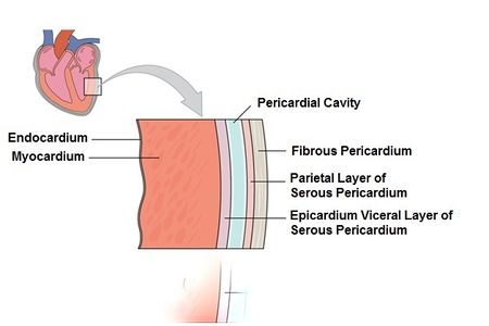
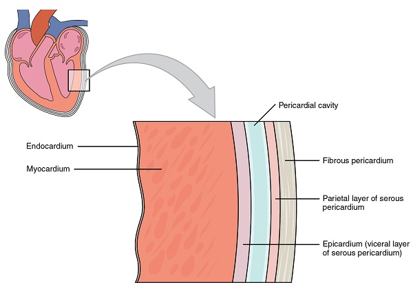
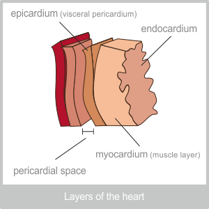
The heart is located in the thoracic cavity medial to the lungs and posterior to the sternum. Pericardium is the outer covering of the heart.
Anatomy Of The Human Heart Physiopedia
The pacemaker device was not implanted but its wire was connected to the patients heart.
Which layer of the heart contains the pacemaker. The chamber heart has a mature type of ECG. The space between the two layers is called pericardial cavity or pericardial space and it contains a thin film of fluid. The endocardium contains smooth muscle.
The SA node is responsible for setting the. Purkinje tissue or subendocardial branches are located in the inner ventricular walls of the heart just beneath the endocardium in a space called the subendocardium. Atrioventricular node AV node.
The impulse then goes to the AV node. It is also known as visceral pericardium as it forms the inner layer of the pericardium. The sino-atrial node or the right wall of the atrium present near the opening of superior vena cava contains the pacemaker.
Click to see full answer. Parietal pericardium forms a strong protective sac for the heart. B The basic configuration of slow conducting pacemaker and fast conducting chamber components is found in the adult fish and human heart.
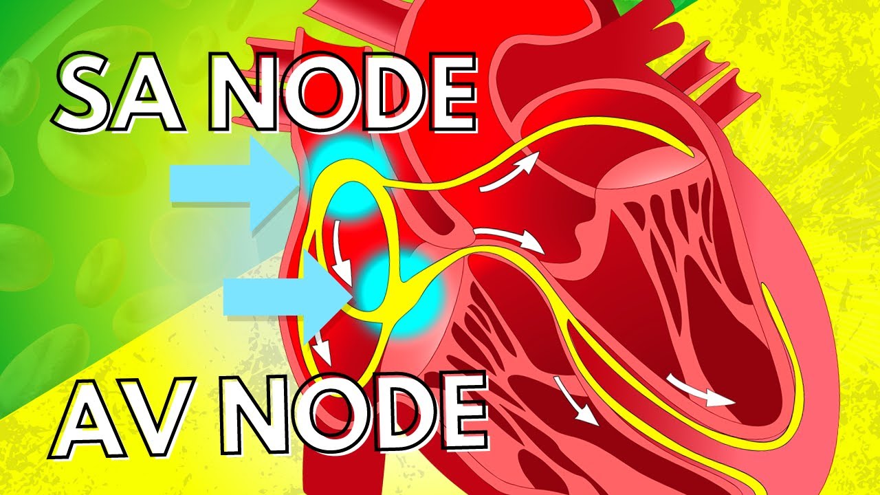
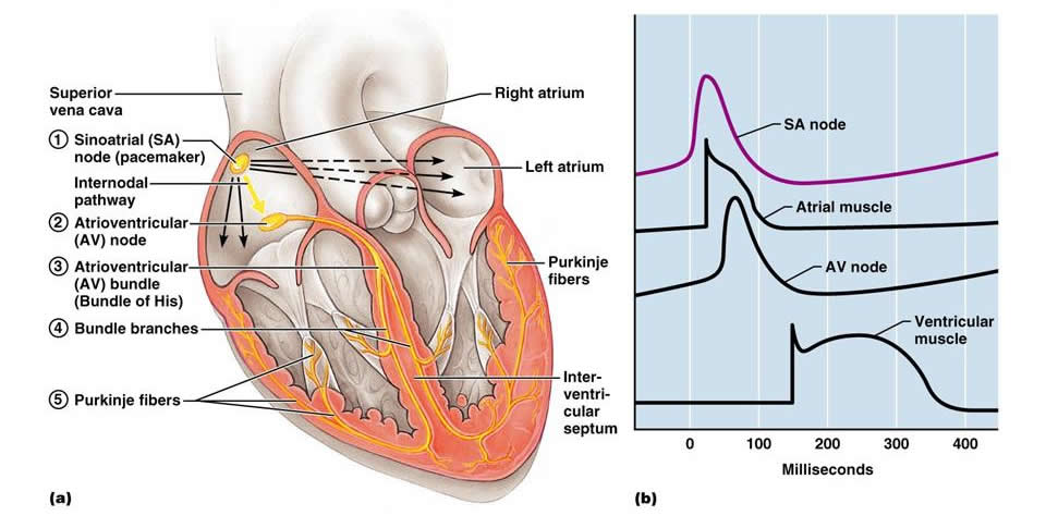
The pacemaker of the heart is the sinoatrial node SA node. The pacemaking signal generated in the sinoatrial node travels through the right atrium to the atrioventricular node along the Bundle of His and through bundle branches to cause contraction of the heart muscle. Between the left atrium and the right ventricle within the atrial septum.
Sinoatrial node SA node. The epicardium is composed primarily of loose connective tissue including elastic fibers and adipose tissue. This sa node is called the hearts natural pacemaker.
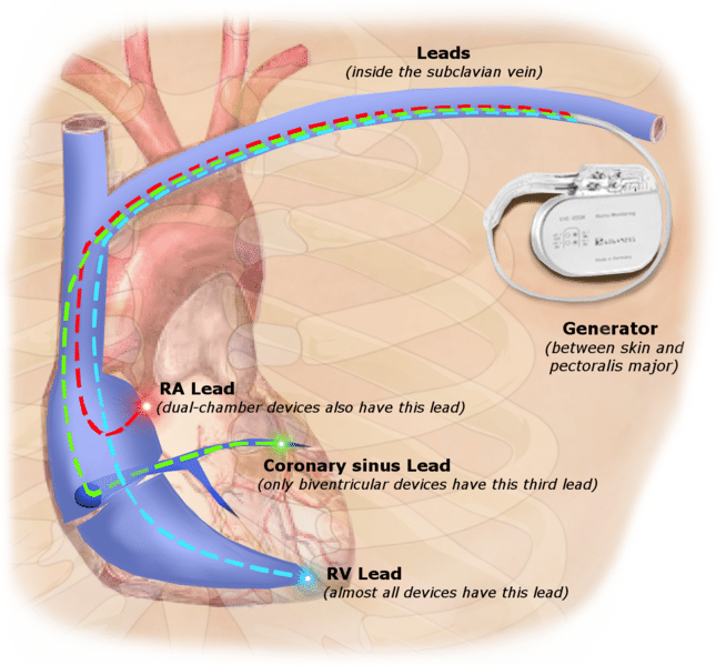
A cardiac impulse is transmitted through a specific pathway of modified cardiac tissue within the heart. The pacemaker of the heart is the sinoatrial SA node which is located in the upper right atrium just below the entrance of the superior vena cava. A pacemaker can pace the hearts upper chambers the atria the lower chambers the ventricles or both.
What is the pacemaker of the heart. This signal stimulates contraction first of the. It is up of two layers.
The endocardium contains abundant adipose tissue b. The endocardium is a thin protective layer on the inside of the heart. The endocardium is layered c.
This electrical signal begins in the sinoatrial SA node located at the top of the hearts upper right chamber the right atrium. Which layer of the heart contains the pacemaker. Pacemakers may also be used to stop the heart from triggering impulses or from sending extra impulses.
Which part of the cardiac conduction system is known as the pacemaker of the heart. The first pacemaker the result of long arduous research was used in a patient in 1958. Which of the following is not true regarding the endocardium.
It is made of smooth slippery endothelial cells which prevent blood from sticking to the inside of the heart and forming deadly blood clots. C The sinus node san forms in the sinus venosus the AVN avn and atrioventricular junction avj in the atrioventricular canal. Pacemakers are available to correct virtually any form of arrhythmia.
SA node Which part of the cardiac conduction system is located in the walls of the ventricles and cause the ventricles. The pacemaker is located in the wall of the right atrium both ventricles and septum. The pacemaker is located in the wall of the right atrium both ventricles and septum.
The endocardium contains blood vessels d. Bundle of His d. The pacemaker cells of the heart with location are.
Wall of the right atrium near the opening of the superior vena cava. Right bundle branch e. The Purkinje fibers pɜːrˈkɪndʒi pur-KIN-jee.
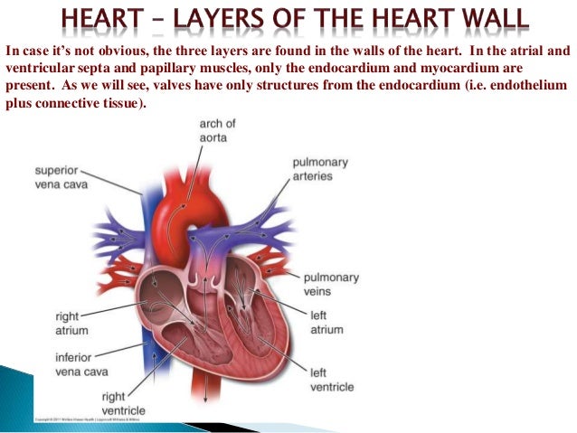
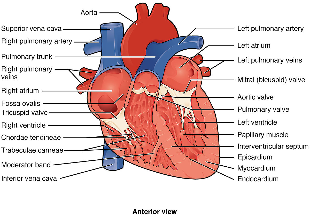
The Purkinje fibers are specialized conducting fibers. Serous fluid secreted by both the parietal and visceral layers of the pericardium and released into the pericardial cavity lubricates the membranes and facilitates the almost frictionless movement of. Chambers of the Heart.
It takes in deoxygenated blood through the veins and delivers it to the lungs for oxygenation before pumping it into the various arteries which provide oxygen and nutrients to body tissues by transporting the blood throughout the body. The pacemaker itself was so large that it had to be carted around in a grocery store cart. Pacemakers can help pace the heart in cases of slow heart rate fast and slow heart rate or a blockage in the hearts electrical system.
Epicardium epi- cardium is the outer layer of the heart wall.

Heart Anatomy Pathology And Components Of The Cvs Lecturio

Heart Amboss
301notes5
Descripition Of Heart And Purkinje Fibres
Heart Anatomy Pathology And Components Of The Cvs Lecturio

Pericardium Wikipedia
Trabeculae Carneae Wikipedia
Heart Anatomy Pathology And Components Of The Cvs Lecturio
The Heart The Function Of The Heart Explained By The Torbay Acupuncture Centre
Conducting System Of The Heart Bundle Of His Sa Node Teachmeanatomy
Where Are The Pacemaker Cells Located In The Heart Socratic
Pacemaker Insertion Johns Hopkins Medicine
.ashx?h=244&la=en&mh=360&mw=520&w=220&hash=9FB84D587FA19BE63DDFD9823A53DEEF012B2EAA)

Cardiac Pacemaker Wikipedia

Pericardium Wikipedia